Key to Alzheimer’s Disease in Simple Amino Acid?

For over a decade, big pharmaceutical companies have invested billions in Alzheimer’s disease drug trials without making significant progress.

However, a potential neuroprotective compound with promising early-stage results might be found in our everyday diet.

Dr. Paul Cox may have discovered this after investigating high rates of ALS and Alzheimer’s-like symptoms in Guam during the 1990s. He found that cyanobacteria, which produce a toxin called BMAA, were contaminating trees on the island. The trees’ seeds, eaten by flying fox bats, became a source of BMAA for the local population, who hunted the bats for food. This toxin was linked to widespread neurodegenerative diseases among the locals.

In 2003, Dr. Cox revealed that cyanobacteria could be a risk factor for these diseases, though not necessarily the cause of Alzheimer’s. To study BMAA’s toxicology, he conducted trials at the Brain Chemistry Labs at the Institute for Ethnomedicine in Jackson. His research showed that the neurotoxic effects of BMAA were reduced by 85% when combined with the amino acid L-serine.

L-serine, found in eggs, meat, edamame, tofu, seaweed, and sweet potatoes, is a non-essential amino acid in our diet. The protective effects observed in monkeys prompted Dr. Cox to initiate clinical trials with the FDA to explore L-serine as a potential Alzheimer’s treatment.

Interestingly, Dr. Cox is an ethnobotanist, not a neurologist. His research in Okinawa, a region known for its longevity, revealed that residents of Ogimi Village consumed about 400% more L-serine than the average American. This, combined with his lab data, gives Dr. Cox confidence that his clinical trials with Alzheimer’s patients supplementing with L-serine could yield effective results, potentially leading to a simple dietary treatment for the disease.

Scientists Grow Micro-Diamonds in 15 Minutes

In South Korea, chemists have recently developed a method to grow artificial micro-diamonds in minutes instead of days.

Remarkably, this technique does not require high temperatures or intense pressure and creates diamonds “from scratch.” This breakthrough has the potential to revolutionize the diamond industry by providing an unlimited supply of micro-diamonds for polishing and cutting applications.

Typically, gemstones form under intense heat, pressure, natural catalysts, or a combination of these factors. Artificially manufacturing diamonds has traditionally required significant amounts of heat and pressure. However, Rodney Ruoff, a physical chemist at the Institute for Basic Science in South Korea, has found a way to eliminate these requirements.

Ruoff and his colleagues used only graphene, silicon, gallium, iron, and nickel—foregoing the need for gigapascals of pressure and temperatures as high as 2,700 degrees Fahrenheit (1,500 degrees Celsius).

Ruoff’s method began with gallium, which had previously been shown to catalyze the formation of graphene. Graphene is pure carbon, similar to a diamond, but with a straight and flat chemical structure, unlike diamond’s tetrahedral formation.

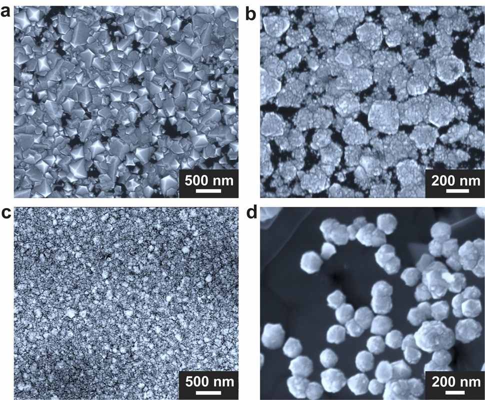
The most expensive component of the experiment was a home-built chamber designed by co-author Won Kyung Seong. This chamber housed the gallium-nickel-iron mixture in a graphene crucible and withstood the introduction of hot methane gas. When all these elements combined, along with a pinch of silicon, diamonds formed within 15 minutes at sea-level pressures inside the chamber.

The precise chemical details of the experiment’s success are still not fully understood, and it will likely be a few years before scientists can confirm whether Ruoff’s process can produce diamonds suitable for jewelry. The diamonds described in their study were thousands of times smaller than lab-grown diamonds typically used for jewelry.

However, the film of micro-diamonds could replace larger diamonds for purposes such as being crushed into powder. Diamond is used for many different cutting and grinding tools and is used from construction to cutting and polishing diamonds themselves.

Type 2 Diabetes Cured by Stem Cells

Stem cells are increasingly being used in a variety of medical treatments, but a recent breakthrough from Shanghai suggests the best is yet to come.

A senior who had suffered from type-2 diabetes for 25 years hasn’t taken insulin for 33 months after receiving a regenerative islet cell transplantation.

Type-2 diabetes, often developed due to poor diet and lifestyle choices, is one of the most prevalent non-communicable diseases globally. China is particularly affected, with 140 million people suffering from diabetes-related complications such as kidney problems, blindness, amputation, and cardiovascular issues.

However, this new breakthrough, the result of 10 years of research and testing, might revolutionize the treatment of diabetes.

Yin Hao, a leading researcher and director of Shanghai Changzheng Hospital’s Organ Transplant Center, explained that they took the patient’s own peripheral blood mononuclear cells and reprogrammed them into pluripotent stem cells, which were then injected into the pancreas.

“Our technology has matured and it has pushed boundaries in the field of regenerative medicine for the treatment of diabetes,” Yin told China Daily. His team conducted the research with scientists from the Center for Molecular Cell Science at the Chinese Academy of Sciences.

Current transplant treatments for type-2 diabetes are limited by a shortage of donor cells and the complexity of pancreatic islet cell isolation technology. Pancreatic islet cells are the main insulin producers in the body, and the patient’s cells were almost completely inhibited. He relied on multiple daily insulin injections and had also undergone a kidney transplant.

After receiving the manufactured stem cells in 2021, he gradually stopped needing external insulin over 11 weeks, and his diabetes seemed largely cured.

“Follow-up examinations showed that the patient’s pancreatic islet function was effectively restored, and his renal function was within normal range,” Yin said. “Such results suggested that the treatment can avoid the progression of diabetic complications.”

The study was published in Cell Discovery on April 30th. Future research, the authors wrote, should explore pharmacological options that could provide off-the-shelf equivalents for islet transplantation.

Blood Test for Strokes

Stroke is the leading cause of disability worldwide and the second leading cause of death. Early intervention can mitigate severe consequences, and a new diagnostic tool could significantly improve outcomes.

Researchers at Brigham and Women’s Hospital have developed a test that uses blood-based biomarkers and a clinical score to accurately identify patients with large vessel occlusion (LVO) strokes.

“We have developed an accessible tool that could help ensure more stroke patients receive timely, critical care,” said Joshua Bernstock, MD, PhD, a clinical fellow in the Department of Neurosurgery at the hospital.

Most strokes are ischemic, caused by obstructed blood flow to the brain. LVO strokes, a severe form of ischemic stroke, occur when a major brain artery is blocked. These require immediate treatment with mechanical thrombectomy, a procedure to remove the blockage, to prevent brain cell death and severe disability.

“Mechanical thrombectomy can restore patients to their pre-stroke condition,” explained Dr. Bernstock.

However, symptoms of brain bleeds can mimic those of LVO strokes, requiring different treatments. The new test distinguishes between the two conditions in the field.

The research team targeted two proteins in capillary blood: glial fibrillary acidic protein (GFAP), associated with brain bleeds and traumatic brain injury, and D-dimer. Their study demonstrated that combining these biomarkers with field assessment scores (stroke triage for emergency destination, or FAST-ED) can identify LVO strokes and rule out brain bleeds.

“The sooner this intervention occurs, the better the patient’s outcome,” said Bernstock. The study, published in the journal Stroke, showed that the test identified LVO strokes with 93% specificity and 81% sensitivity within six hours of symptom onset. It also ruled out all patients with brain bleeds.

This tool could be especially valuable in low- and middle-income countries where advanced imaging is unavailable. It might also aid in diagnosing traumatic brain injuries. The team is conducting a prospective trial to test the tool’s performance in ambulances and has designed an interventional trial to speed up stroke triage by bypassing standard imaging.

“In stroke care, time is brain,” Bernstock emphasized. “The sooner a patient is on the right care pathway, the better their outcome will be. Our prehospital diagnostic tool is set to be transformative.”

Experimental Cancer Treatment Gives Mom a 2nd Chance

After less than a year of undergoing treatment with a new cancer drug, a young woman saw her tumor disappear, easing concerns about her ability to have more children.

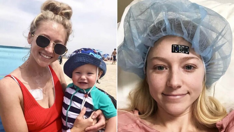
Kelly Spill’s life took a difficult turn after giving birth to her first baby when she was diagnosed with cancer. Experiencing symptoms like fatigue, bleeding, weight loss, and loss of appetite, she suspected cancer despite doctors initially attributing her symptoms to childbirth.

Tests confirmed stage-3 colorectal cancer, leading Spill to seek treatment at Memorial Sloan Kettering Cancer Center. Conventional treatment options posed a risk to her fertility, a concern as she hoped for a larger family.

However, just before starting chemotherapy, Spill became eligible for a clinical trial testing a new drug, dostarlimab. She received weekly injections for six months.

The treatment proved effective, with her tumor significantly shrinking after four treatments and disappearing completely by the ninth. Spill cautiously considered expanding her family and eventually gave birth to a healthy baby girl.

Today, Spill remains cancer-free, her journey showcasing the potential of innovative cancer therapies.